Are nuclear masks all you need for improved out-of-domain generalisation? A closer look at cancer classification in histopathology
NeurIPS · 2024
Summary
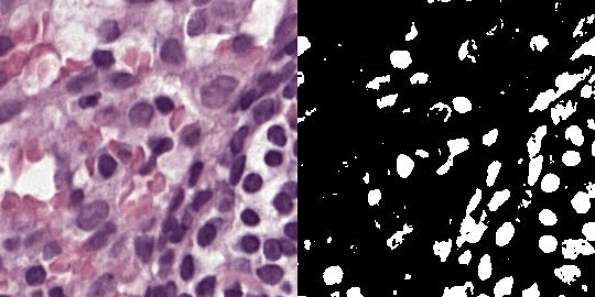
Models trained on data from one hospital often struggle when used on data from other hospitals. We guide the training of a standard image classifier using nuclear masks, helping it focus more on nuclear shape and arrangement. This approach improves accuracy and robustness across hospitals, and the model does not need masks during testing.
The problem
One of the biggest hurdles in medical AI is that a cancer-detecting model trained at Hospital A often fails when tested at Hospital B.
Why does this happen? To look at tissue under a microscope, it must be sliced very thin and stained with chemicals—most commonly haematoxylin and eosin (H&E), which turns cell nuclei purple and surrounding tissue pink. But hospitals use different scanners, different staining procedures, and even different batches of chemicals.
To a human pathologist, these visual differences are minor. But a neural network will often lazily memorize the specific “look” of its training hospital. When the image appearance changes, the model breaks.
Our paper asks a simple question: Can we force a cancer classifier to stop looking at hospital-specific colors, and instead focus on the biological structures that actually matter?
The idea: Shape over color
While the exact shade of pink or purple might change depending on the hospital, the underlying biology of cancer does not. Cancer notoriously alters the shape, size, and crowding of cell nuclei. Pathologists rely heavily on these structural changes to diagnose the disease.
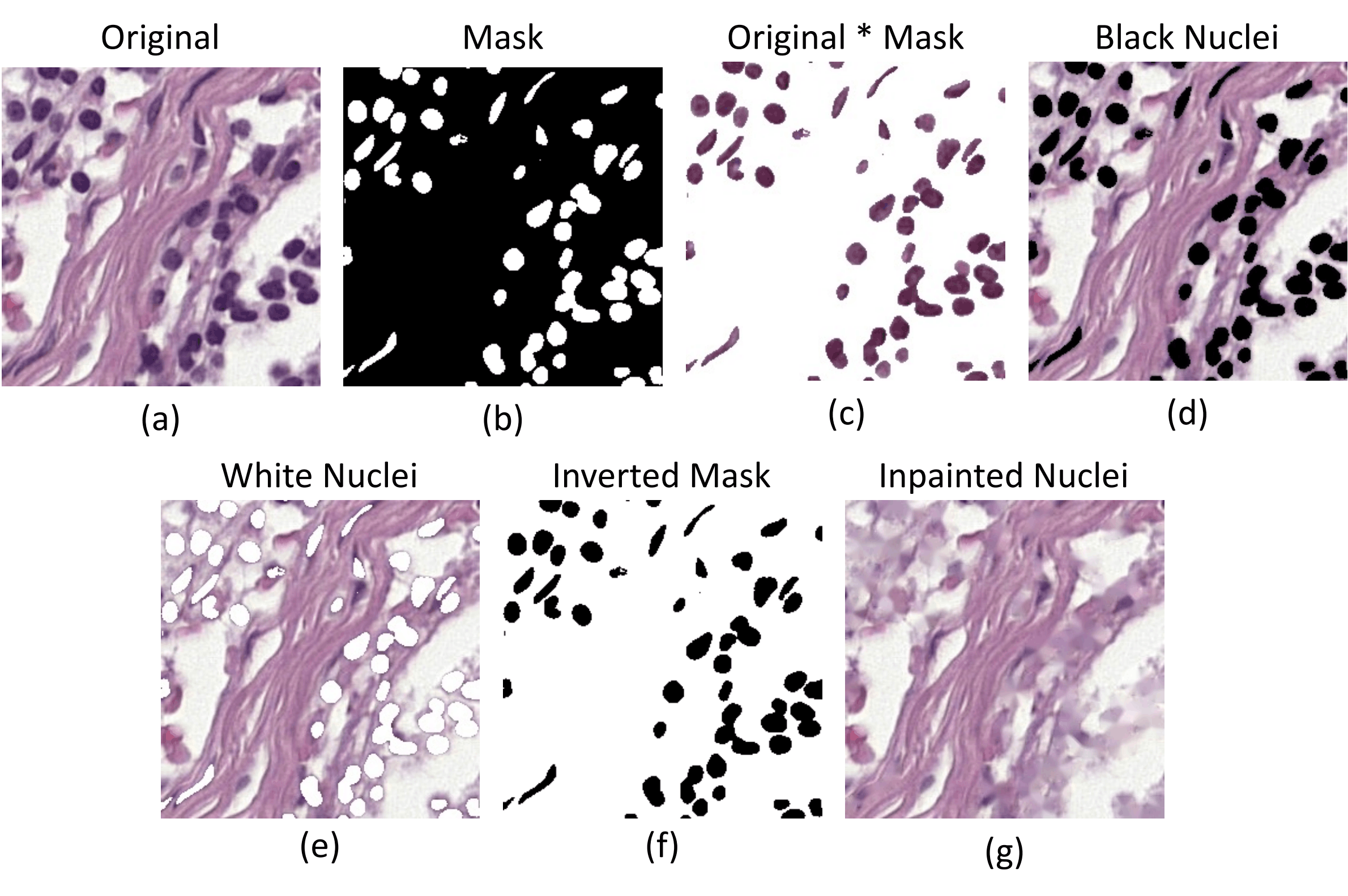
To see if we could isolate this structural signal, we generated simple binary masks: black-and-white images where the nuclei are white and absolutely everything else is black.
This led to a turning point in the project. We found that a model trained only on these stripped-down masks could still accurately classify cancer—and crucially, it generalised beautifully to data from other hospitals. The shape and arrangement of the nuclei contained all the information we needed.
But there was a catch. If we deployed a mask-only model in the real world, hospitals would have to run a separate segmentation algorithm on every single image before diagnosing it. Our goal was to use these masks only as a teacher during training, so the model could learn from them but wouldn’t need them in practice.
How the method works
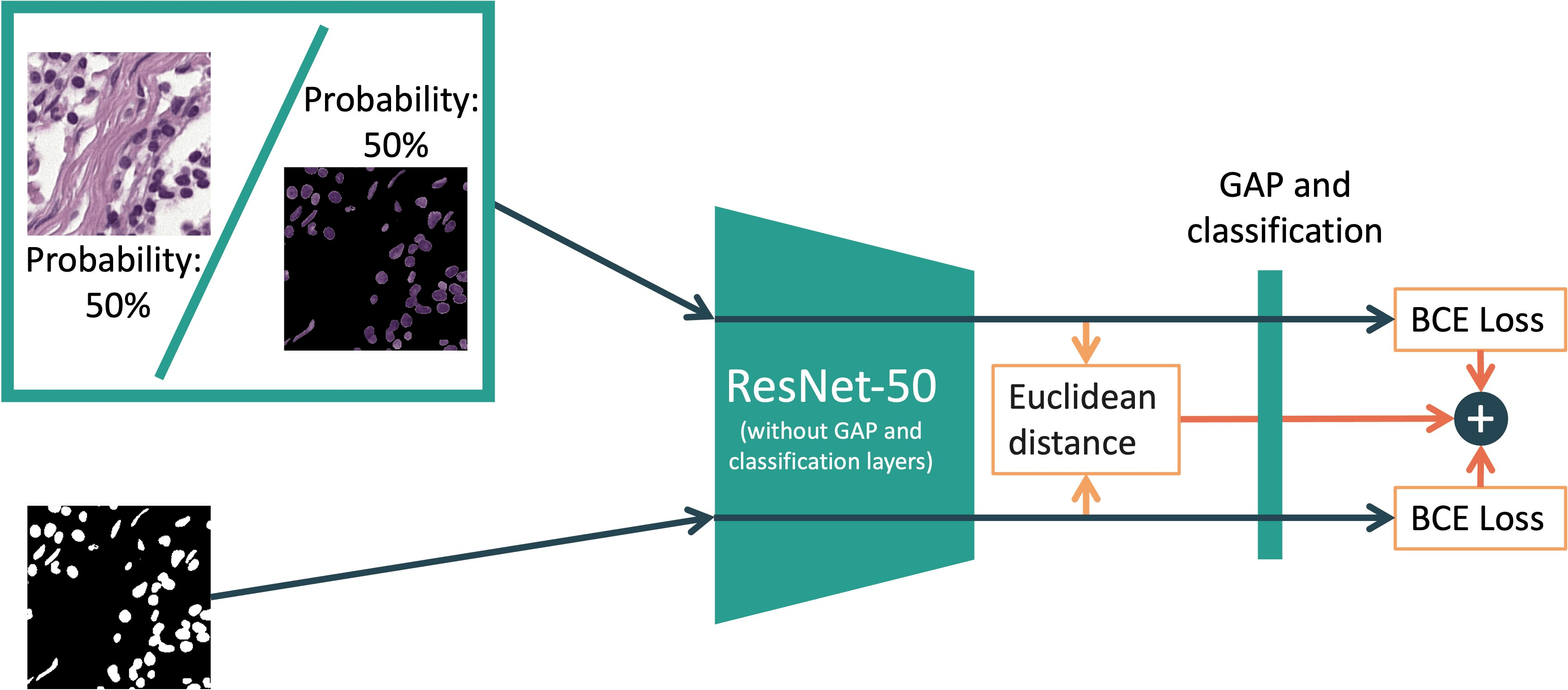
We designed a two-branch training method using a standard ResNet-50. During training, the model looks at two aligned inputs simultaneously:
- The full-color H&E image (and occasionally the image multiplied by its mask).
- The black-and-white nuclear mask.
The model is trained to classify both. But here is where the magic happens: we explicitly minimise the Euclidean distance between the internal feature maps of both inputs (right before global average pooling).
In plain English: We mathematically force the model’s understanding of the full-color image to match its understanding of the black-and-white mask. It’s like giving a student a highly detailed photograph and a basic sketch, and training them to focus only on the structural features they both share.
To make this alignment easier for the model to learn, we sometimes feed the image branch a “bridged” input: the original image multiplied by the mask, which keeps the nuclear colors but blacks out the background.
Evaluation
We trained models on one medical centre (hospital) from CAMELYON17 at a time and evaluated them on the other four centres. We then tested the final models on two external datasets:
- BCSS, containing primary breast-cancer tissue from TCGA; and
- OCELOT, containing primary tumours from six organs.
The external datasets were held back until the final evaluation. We compared against ordinary empirical risk minimisation (ERM), Macenko stain normalisation, RandStainNA, RSC, L2D, DDCA, and a model initialised from HoVer-Net weights. Among the baselines, L2D performed best, while ERM (with or without Macenko normalisation) is the most commonly used method in the histopathology community.
| Method | CAMELYON17 | BCSS | OCELOT |
|---|---|---|---|
| ERM-Aug | 85.5 ± 1.8 | 71.7 ± 2.3 | 65.6 ± 2.1 |
| L2D-Aug | 89.1 ± 1.1 | 75.1 ± 1.3 | 69.1 ± 1.1 |
| Ours-Aug | 91.8 ± 0.9 | 78.8 ± 1.6 | 70.6 ± 1.0 |
With photometric augmentation, our method reached average accuracies of 91.8% on CAMELYON17, 78.8% on BCSS, and 70.6% on OCELOT. The corresponding L2D results were 89.1%, 75.1%, and 69.1%; the ordinary augmented baseline reached 85.5%, 71.7%, and 65.6%.
The important result is not simply that nuclear guidance replaced colour augmentation. The two were complementary: conventional augmentation broadened the visual variation seen during training, while nuclear alignment encouraged the model to preserve a biologically meaningful signal across that variation.
Robustness beyond hospital shifts
Because our model relies on structure rather than superficial color cues, we wanted to see if it was physically harder to break.
First, we tested common image corruptions (like blur, heavy JPEG compression, and digital noise). Models trained with our nuclear-guided method degraded much more slowly than standard models.
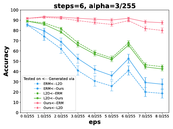
Second, we tried to deliberately trick the AI using adversarial attacks (specifically, projected-gradient descent). Our models remained accurate even when hit with heavy digital perturbations that caused standard models (like ERM and L2D) to completely fail.
Interestingly, when we used standard models to generate adversarial images and fed them to our model, our model wasn’t easily fooled. But when we generated attacks against our model and fed them to the standard baselines, those models broke down. My reasoning is that since our model is trained to ignore the background and focus on nuclei, an attacker practically has to alter the nuclear structure itself to trick it! Although this reasoning is consistent with the results, I need to point out that the experiment alone does not prove the mechanism.
Did the model really focus on nuclei?
We used several ablations to test whether the method had changed what the model relied on:
- When we removed the non-nuclear background, performance remained high.
- When we subtracted mask-like features from the image representation, our model suffered the largest drop.
- When nuclei were inpainted away, our model predicted almost no tumour tiles.
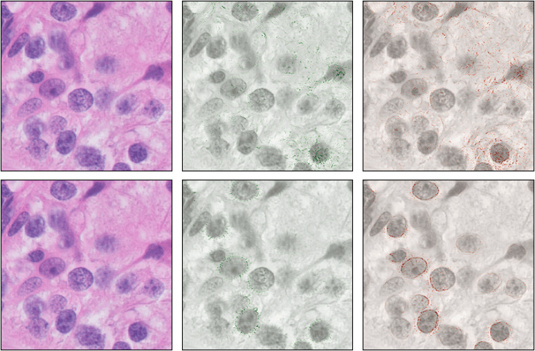
- Integrated Gradients highlighted nuclear regions more clearly for our method than for the ordinary augmented baseline.
Limitations
The main experiments study one binary classification problem and use ResNet-50 as the primary architecture. We included a preliminary ViT-Tiny evaluation, but larger transformer studies were outside the scope of the paper. The method also depends on having usable nuclear masks during training, even though it does not need them at inference time.
The claim should therefore remain scoped: nuclear masks were highly informative for the cancer-classification tasks and datasets studied here. Other histopathology tasks may depend more heavily on tissue morphology or non-nuclear context.
Want the behind-the-scenes story?
Academic papers only tell half the story. If you want to read about the messy reality of how this research actually came together—including the failed early experiments with Canny edge detectors, the struggle to stabilise the training loss, and the incredibly lucky misunderstanding that led to our favourite adversarial robustness result—check out the companion blog post:
Read the story: What happens when a cancer model can see only cell nuclei?
Contributions
- A method that uses nuclear segmentation masks, only during training, to steer a classifier towards nuclear morphology and organisation & does not require nuclear masks at inference time.
- A comparison with popular stain normalisation, data augmentation, and single-domain generalisation methods across CAMELYON17, BCSS, and OCELOT datasets.
- Robustness experiments covering common image corruptions, adversarial attacks and cross-model attack transfer. Many ablation experiments.
Citation
Tomar, D., Binder, A., and Kleppe, A. (2024). Are nuclear masks all you need for improved out-of-domain generalisation? A closer look at cancer classification in histopathology. Advances in Neural Information Processing Systems, 37, 43499–43532.
@inproceedings{tomar2024nuclear,
  title     = {Are nuclear masks all you need for improved out-of-domain generalisation? A closer look at cancer classification in histopathology},
  author    = {Tomar, Dhananjay and Binder, Alexander and Kleppe, Andreas},
  booktitle = {Advances in Neural Information Processing Systems},
  volume    = {37},
  pages     = {43499--43532},
  year      = {2024},
  doi       = {10.52202/079017-1378}
}